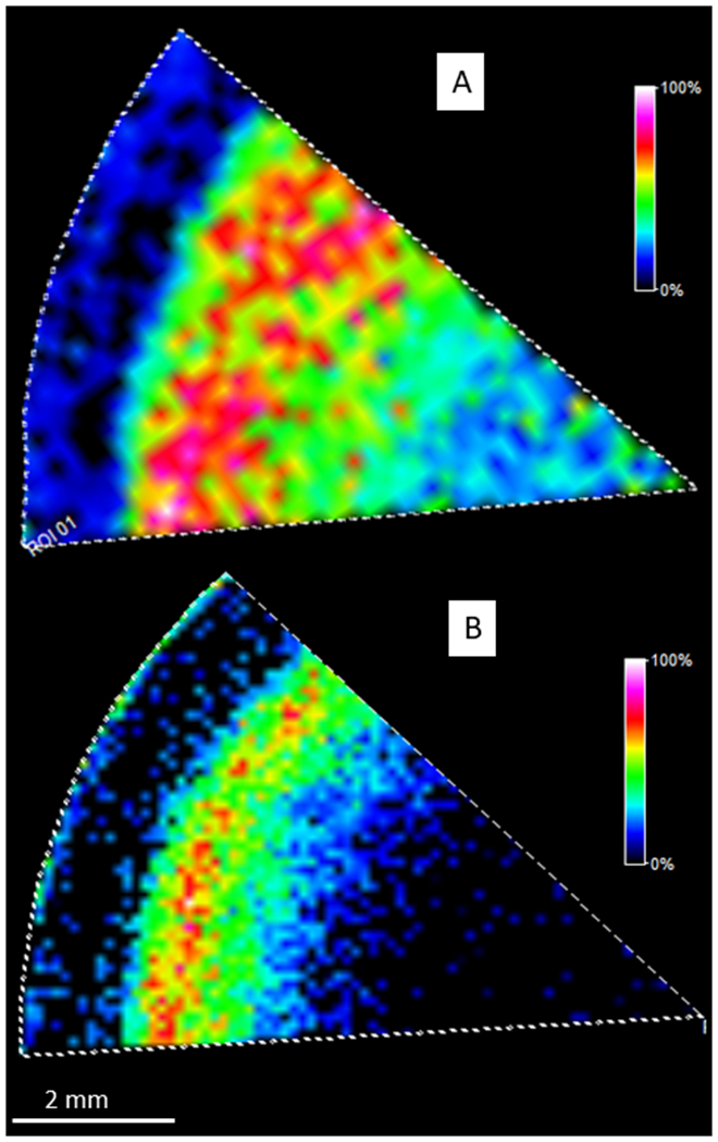
Figure 6.

MALDI imaging of myristoylated filensin peptides. A: MALDI imaging of myristoylated 432-434 (m/z 471.3541±10 ppm) after a combination of LysC and Trypsin digestion. B: MALDI imaging of myristoylated 432-440 (m/z 1146.7133±10 ppm) after GluC digestion. Scale bar: 2 mm. Both results indicate that myristoylation starts at a normalized lens distance of 0.87.
