Figure 4.
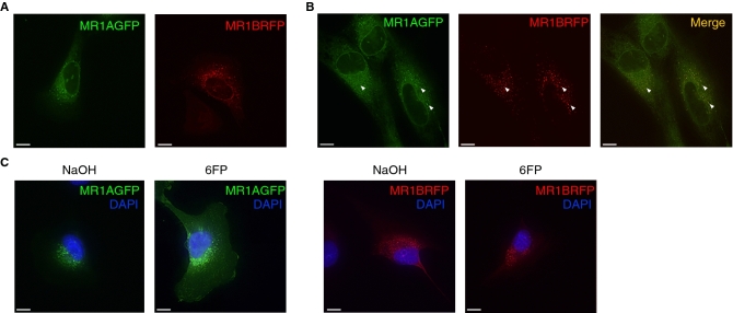
MR1A and MR1B reside in the same intracellular compartments. (A) Beas2B:MR1_KO cells were transfected with plasmids encoding either MR1AGFP or MR1BRFP. Live cell imaging was performed using a CoreDV microscope to detect intracellular MR1A (left) or MR1B (right). Scale bars represent 10 µm. (B) MR1A-overexpressing Beas2B cells were transfected with a plasmid encoding MR1BRFP and live cell imaging was performed as described in (A) to determine the subcellular localization of MR1A and MR1B. (left) MR1A alone, (middle) MR1B alone, right (merge). (C) Beas2B:MR1_KO cells were transfected with plasmids encoding either MR1AGFP or MR1BRFP, and subsequently treated with 50 µm 6FP or NaOH for 16 h. Cells were fixed for 20 min with 4% PFA and imaging was performed using a CoreDV microscope to determine localization of MR1A or MR1B with or without 6FP. DAPI nuclear stain was used to identify the nucleus. Scale bars represent 10 µm. Experiments were repeated 2–4 times with similar results.

