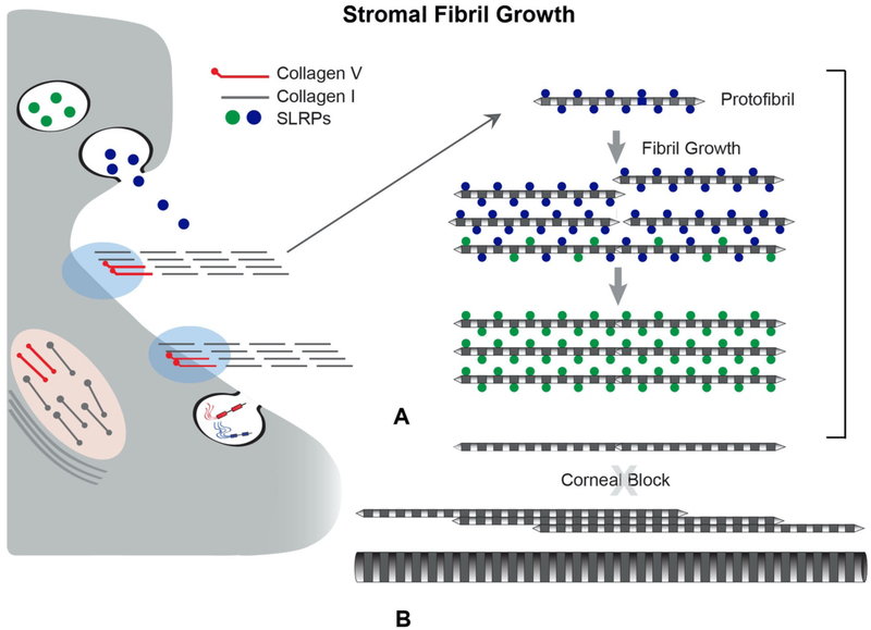
Fig 9. SLRPs Regulate Lateral Fibril Growth.
SLRPs regulate linear and lateral stromal collagen fibril growth by binding to fibril surfaces. (A) Newly assembled stromal fibrils (protofibrils) are deposited into the matrix where they are stabilized via interactions with SLRPs. After deposition into the stromal matrix, protofibrils mature by a process of fibril growth. Bound SLRPs mediate and controlled fibrillar interactions resulting in coordinated growth of mature stromal fibrils. In the stroma there is a a tissue-specific regulation of lateral fibril growth. Diameter and packing are rigidly regulated for corneal transparency. (B) In most tissues there is a robust lateral growth resulting in heterogeneous populations of large diameter fibrils this is blocked in the stroma. Changes in fibril stabilization necessary for growth can result from processing, turnover, and/or displacement of SLRPs. SLRPs affect fibril diameter and spacing in the corneal stroma. When SLRPs are absent, as in null mice or in gene mutations in human patients, fibril structure, organization and spacing are impacted, compromising transparency. Also, the block to lateral fibril growth is lost in the stroma when SLPR expression is altered.

