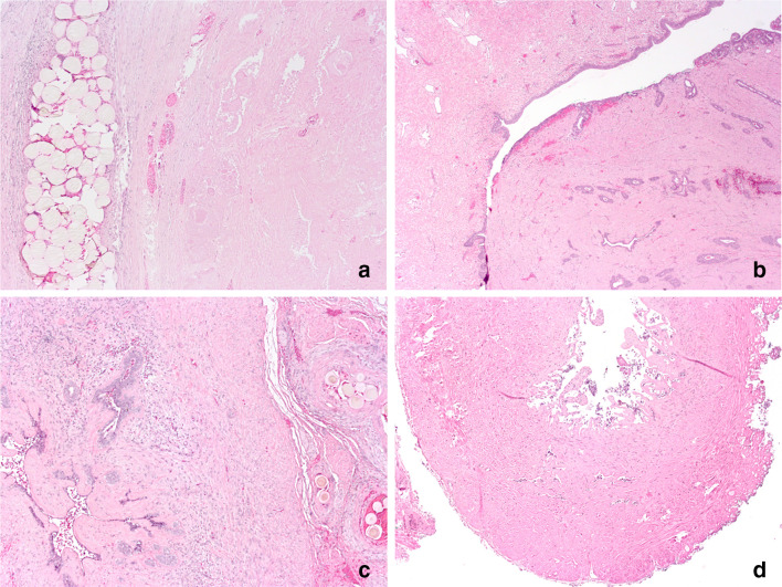
Fig. 2.
Histological findings in patients undergoing radical prostatectomy 6 weeks after prostatic artery embolization. Selected pictures (HE staining) show extensive necrosis next to embolization particles in the central prostatic gland (a, × 50), extensive fibrosis around the verumontanum (b, × 25) and mucosal necrosis and atrophy of the seminal vesicles (c, × 50) and ejaculatory duct (d, × 50)

