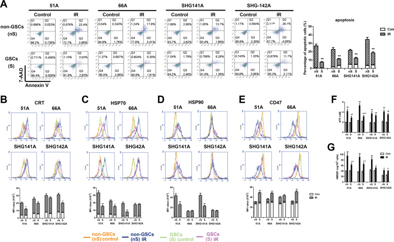
Fig. 1. IR induces less cell apoptosis, immunogenic molecule exposure and release in GSCs compared with non-GSCs.
GSCs and non-GSCs were incubated for 24 h after 10 Gy IR for apoptosis assay and detection of CRT, HSP90, HSP70, CD47, ATP, and HMGB1. a Cell apoptosis was determined by Annexin V and 7-AAD staining. The expressions of CRT (b), HSP70 (c), HSP90 (d), and CD47 (e) on cell surface were measured by flow cytometry. The level of extracellular ATP (f) using a chemiluminescence assay and HMGB1 release (g) in supernatant using an elisa assay were detected. *P < 0.05, **P < 0.01 vs matched non-GSCs, #P < 0.05, ##P < 0.01 vs control.

