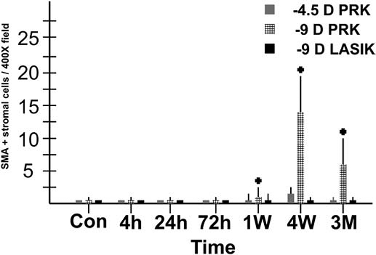
Fig. 6.

Quantitative analysis of alpha-smooth muscle actin+ myofibroblasts at each time point for −4.5D PRK, −9D PRK and −9D LASIK, with 8 corneas in each group at each time point. Note myofibroblasts were much greater in the −9D PRK group, with only a few myofibroblasts detected in corneas that had −4.5D PRK and none detected in the central cornea of eyes that had −9D LASIK. Modified with permission from Mohan et al. Exp. Eye Res. 2003;76:71-87.
