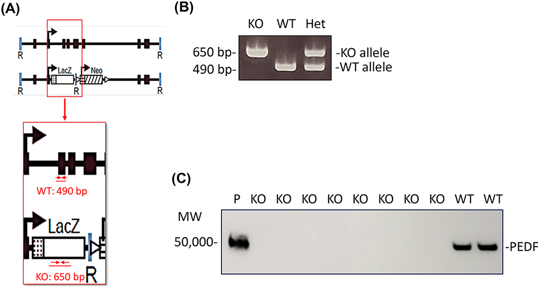
Fig. 2.

(A) Serpinf1 null mice (KO), organization illustrating location of primers for genotyping and expected product size (adapted from Doll et al. 2003). (B) Detection of Serpinf1 gene by genotyping (C) Plasma PEDF protein levels –Western blotting vs anti-PEDF antibody.
