Fig 3:

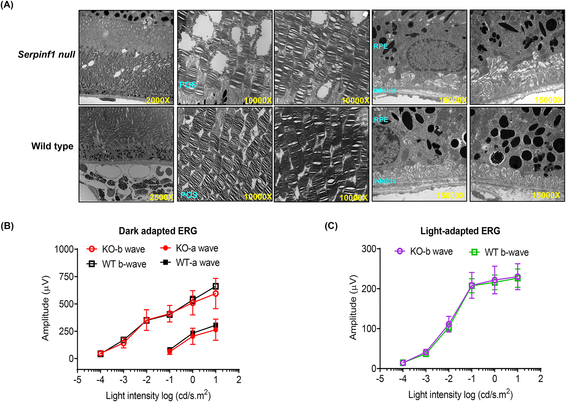
Morphology and visual function of Serpinf1 null (KO) mice and wild type (WT) littermates. (A) Electron microscopic images of KO and WT mice retina sections. Photoreceptor outer segment degeneration is marked by red arrows (B) Electroretinogram (ERG) of dark-adapted WT mice (WT; black squares and lines) and Serpinf1 null (KO; red circles and lines) at 3–6 months of age. Amplitudes of a waves (solid black squares and circles) and b waves (open black squares and circles) are recorded against light stimulus. (C) ERG of light-adapted wild type mice (WT; green squares and lines) and Serpinf1 null mice (KO; purple circles and lines) at 3–6 months of age. (means ± SD, n=5)
