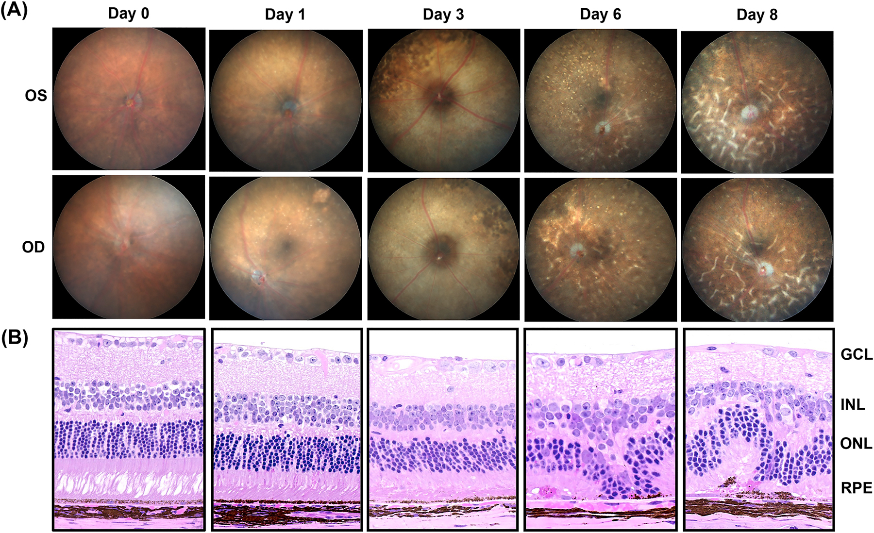
Fig 4:

Progression of retinal damage after NaIO3 treatment in C57BL6/J wild type mice. (A) Follow-up of the retinal damage through fundus microscopy of the mice eyes after 0 (Saline treated), 1, 3, 6 and 8 days post-treatment with NaIO3. (B) Photomicrographs of hematoxylin and eosin stained eye sections of NaIO3-treated mice harvested at different time points. Mice were treated with single intraperitoneal injection of NaIO3 at 30mg/kg body weight. OD=Oculus Dextrus (right eye), OS=Oculus Sinister (left eye), GCL= Ganglion cell layer, INL= Inner nuclear layer, ONL= Outer nuclear layer, RPE= Retinal pigment epithelium. (n=3)
