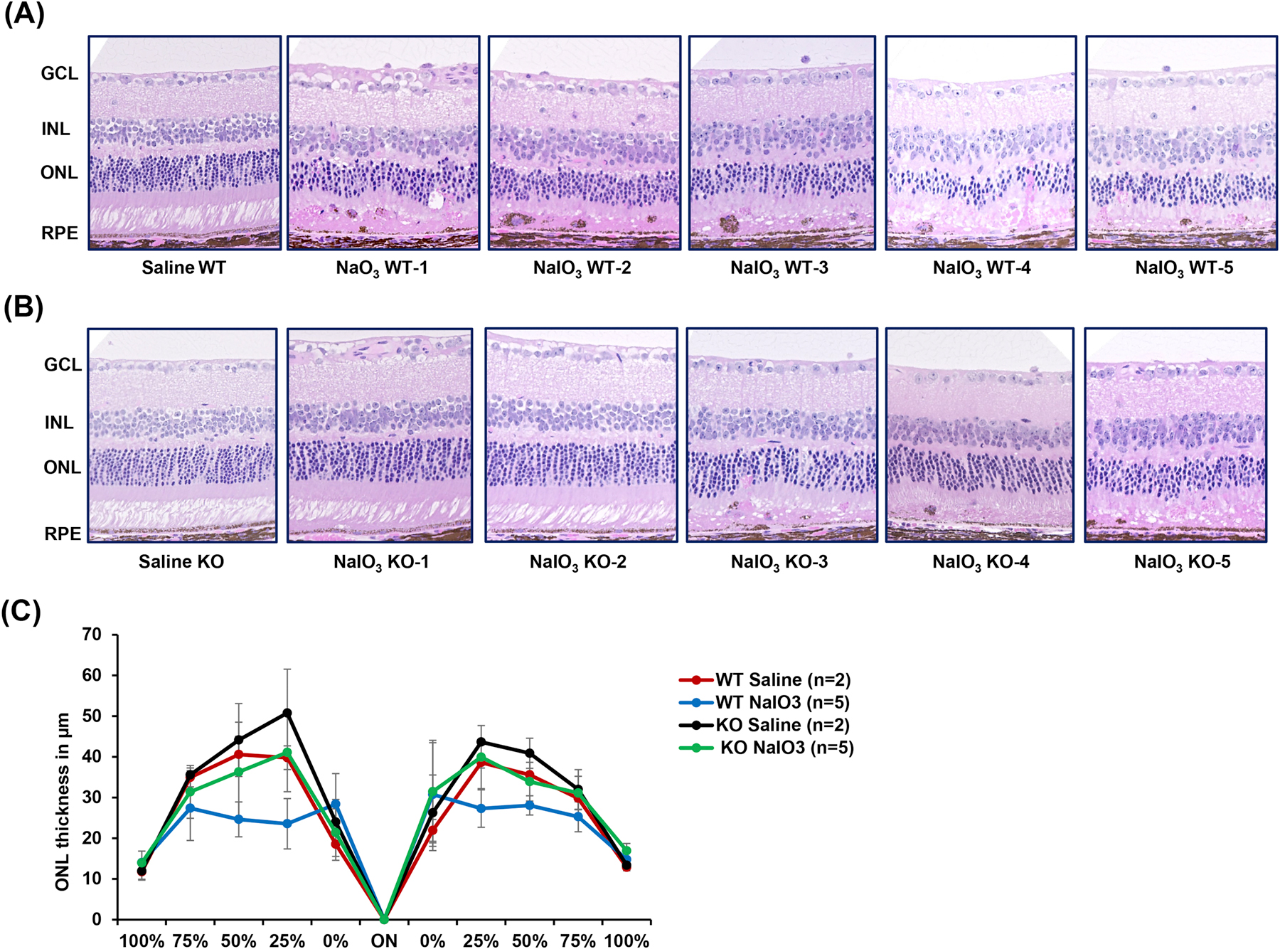
Fig 5:

Histology of retinas of WT and Serpinf1 null mice injected with NaIO3. Serpinf1 null (KO) and WT mice were treated with 30mg/kg body weight of NaIO3 (n=5) or saline (n=2). After 4 days post injection, mice were euthanized and eyes enucleated, fixed, sectioned and stained. (A-B) Microphotographs of retina sections of the saline or NaIO3 treated WT (A) and KO (B) mice stained with hematoxylin and eosin. (B) Spider plot analysis illustrating outer nuclear layer (ONL) thickness in NaIO3 (n = 5) and saline (n=2) treated WT and KO mice.
