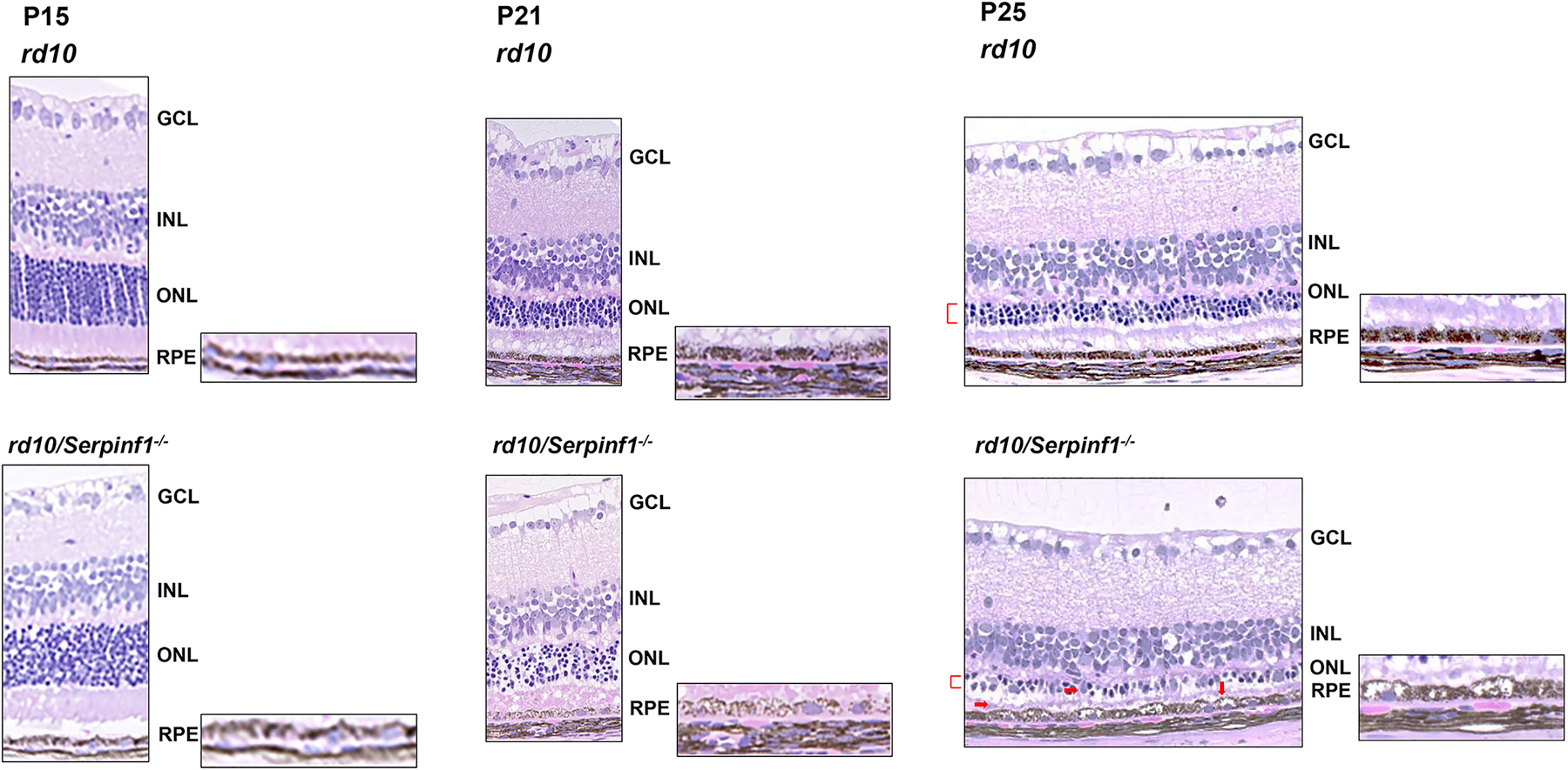
Fig 6:

Histology of retinas of rd10 and rd10/Serpinf1 null mice. Eyes of rd10 and rd10/Serpinf1 null mice and mice at P15, P21 and P25 were enucleated, fixed and stained with hematoxylin and eosin. Representative of images retina sections displaying pathologies associated with PEDF deficiency are shown (red markings). GCL = ganglion cell layer, INL=inner nuclear layer, ONL= Outer nuclear layer, RPE= Retinal pigment epithelium. n=4.
