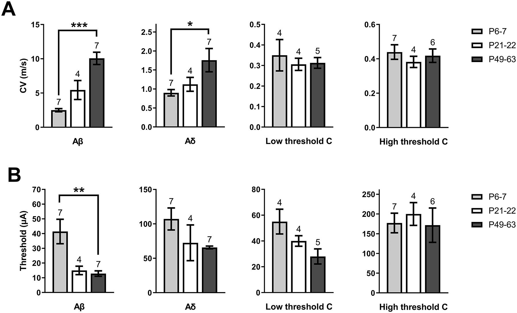
Figure 2: Developmental changes in A-fiber conduction within the mouse sciatic nerve.

A) Mean conduction velocity (CV) of primary afferent types at each age sampled, showing a significantly lower CV in Aβ (H = 13, p < 0.0001; Kruskal-Wallis test) and Aδ fibers (H = 6.15, p = 0.039; Kruskal-Wallis test) during early life compared with adulthood (*p < 0.05, ***p < 0.001; Dunn’s multiple comparisons test; n indicated above bars = number of nerves for all panels). B) Plot of mean stimulus intensity required to elicit a CAP from each fiber type throughout development. The stimulus thresholds of Aβ fibers at P6–7 were higher than in P49–63 mice (H = 10.98, p = 0.0009, Kruskal-Wallis test; **p < 0.01; Dunn’s multiple comparisons test), suggesting lower Aβ fiber excitability in early life.
