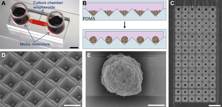
Fig. 1.
Formation of hepatocyte spheroids in microfluidic chambers. A: typical microfluidic device used for cultivation of hepatocyte spheroid contained two media reservoirs connected to a cell culture chamber via transport channels. Scale bar: 5 mm. Media was exchanged every 24 h in this device. Two types of cell culture chambers for cultivation of spheroids were made: 1) 100-μm headspace [small volume three-dimensional (3-D)] and 2) 1,600-μm headspace (large volume 3-D). B: schematic of hepatocytes aggregating into spheroids inside microfluidic chambers containing pyramidal polydimethylsiloxane (PDMS) microwells. The surfaces of microfluidic chambers were precoated with Pluronic to enhance cell-cell interactions. C: a panel of ×4 magnification images stitched together to show spheroid occupancy in a representative culture chamber. Scale bar: 1 mm. D: scanning electron microscopy (SEM) image of an array of hepatocyte spheroids formed in pyramidal PDMS wells collected at ×50 magnification (scale bar: 50 µm). E: SEM image of an individual hepatocyte spheroid imaged at ×500 magnification (scale bar: 500 µm).

