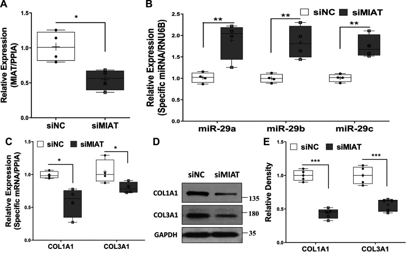
Fig. 4.
A–C: qRT-PCR analysis of myocardial infarction-associated transcript (MIAT; A), miR-29 family (B), or expression of COL1A1 and COL3A1 (C) in rat neonatal primary cardiac fibroblasts after transfection of siRNA negative control (siNC) or MIAT siRNA oligonucleotides (siMIAT) for 48 h (A and B; N = 4) or 72 h (C; N = 4). PPIA, peptidylprolyl isomerase A. D: representative of Western blots of COL1A1 and COL3A1 proteins after 96-h transfection of siNC or siMIAT in rat primary neonatal cardiac fibroblasts with the relative density histograms (E; N = 5). Results were analyzed using unpaired t test and are presented as box-and-whisker plot with P values (*P < 0.05; **P < 0.01, and ***P < 0.001) indicated by the corresponding lines. Thin horizontal lines within boxes are medians, cross symbols are mean values, and whiskers indicate maximum and minimum values.

