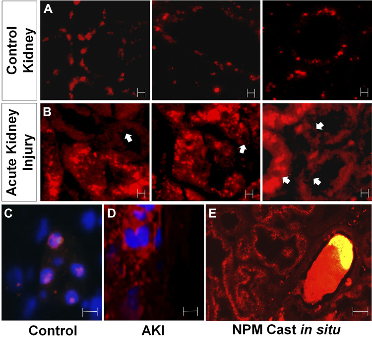
Fig. 8.
Nucleophosmin (NPM) redistribution reflects acute cell injury in human kidney biopsy tissue. A: NPM localizes in a punctate intranuclear pattern in paraffin-fixed human kidney biopsy tissue obtained from three patients with microscopic hematuria caused by thin basement membrane disease but without acute kidney injury (“control”); in control tissue, NPM localized to nuclear regions of proximal tubule cells. Magnification: ×40. B: in contrast, NPM diffusely localized to the cytosol and lumen of kidney tissue in three patients with acute tubular injury (“acute injury”); proximal tubular cells showed NPM distribution in a diffuse cytosolic pattern; arrows show nuclei minimal NPM content. Magnification: ×40. C: high-power view of nuclear NPM staining in a patient with thin basement membrane disease (control) showing NPM-laden nuclei costained with Hoechst dye. D: high-power view of cytosolic NPM staining in a patient with clinical acute kidney injury (AKI) showing distinct distributions for NPM and Hoechst dye. E: an NPM-containing cast trapped inside a collecting tubule lumen. Magnification: ×100. Bars = 5 μm.

