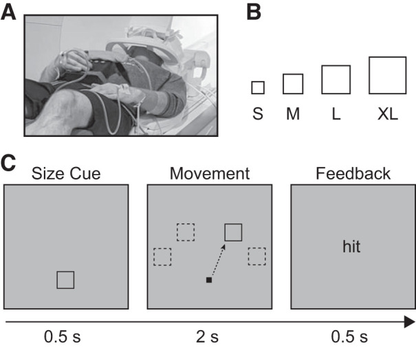
Fig. 1.

Task design. A: participants used an MRI-compatible joystick secured on their lap. Electromyogram (EMG) electrodes were placed on participants’ right and left extensor carpi ulnaris (ECU) muscles. B: target size conditions for the task [small (S): 1.4° × 1.4° visual angle; medium (M): 1.75° × 1.75° visual angle; large (L): 2.1° × 2.1° visual angle; extra large (XL): 2.45° × 2.45° visual angle]. C: each trial began with a size cuing the upcoming movement demand. After the precue, the target appeared in 1 of 4 possible locations (−60°, −30°, 30°, 60° relative to vertical meridian), after which the participants were instructed to use the joystick to move the cursor to the target as quickly and accurately as possible. If the cursor was inside the target 2 s after target presentation, participants received “hit” feedback; otherwise they received “miss” feedback.
