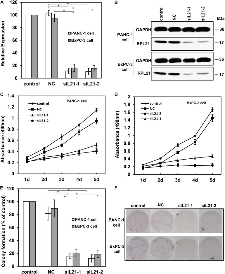
FIGURE 1.
The analysis of cell proliferation on PANC-1 and BxPC-3 cells with siL21-1 and siL21-2 transfection. The quantitative real-time PCR (qPCR) assay (A) and the western blot assay (B) were used to investigate the RNAi effect of siL21-1 and siL21-2 (40 nM, 72 h) in PANC-1 and BxPC-3 cells. GAPDH and β-Actin were used as an internal control for qPCR and western blot analyses, respectively. (C,D) The effect of transfection with siL21-1 and siL21-2 (40 nM) on cell proliferation. The cells were detected by 3-(4, 5-dimethylthiazol-2-yl)-2, 5-diphenyltetrazolium bromide (MTT) assay on each day for 5 consecutive days. (E,F) For colony formation, PANC-1 and BxPC-3 cells were trypsinized and seeded into 35-mm dishes (1,000 cells/dish) at 24 h after transfection. After 8 days, cell colonies with more than 50 cells were dyed with hematoxylin and counted. The control, negative control (NC), siL21-1 and siL21-2 represented the untransfected, Mock-siRNA transfected, siL21-1 and siL21-2 transfected, respectively. Data are presented as the mean ± SD (n = 3). * indicates P < 0.05 compared to the control as determined by the Student’s t-test.

