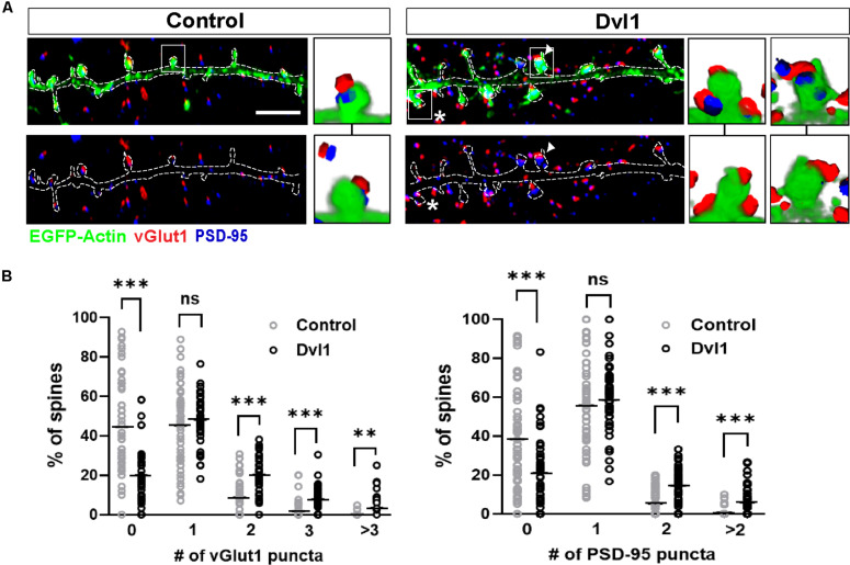
FIGURE 3.
Postsynaptic Dvl1 gain of function increases the innervation of spines and the formation of MIS. (A) Representative images of dendrites expressing EGFP-actin (green) and empty vector (control) or Dvl1-HA and immunostained for vGlut1 (red) and PSD-95 (blue). Dvl1 expression increases the proportion of spines contacted by more than one vGlut1 (asterisks) puncta and spines containing more than one discrete PSD-95 puncta (multiple PSDs; arrowhead) when compared to control neurons. Scale bar = 5μm. Three-dimensional zoomed-in images display front (top) and back (bottom) representations of spines with one or more vGlut1 and PSD95 puncta. (B) Quantification of the distribution of number of vGlut1 and PSD-95 puncta per spine (n = 37–42 cells from 3 independent cultures, **P < 0.01, ***P < 0.001, Mann-Whitney test).

