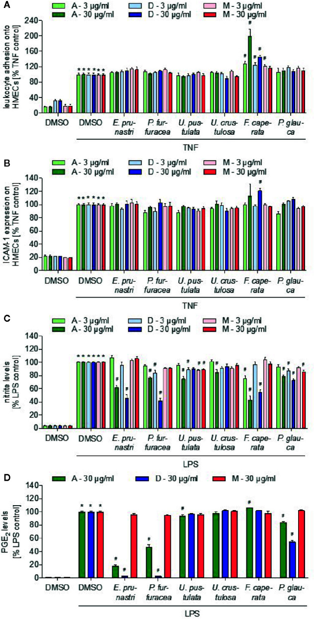
Figure 3.

Effects of lichen extracts on cellular functions. (A, B) HMEC-1 cells were grown to confluence, preincubated with lichen extracts (3 and 30 µg/ml) for 30 min, and activated with TNF (10 ng/ml) for 24 h. (A) For the leukocyte adhesion assay, untreated THP-1 cells were stained with CellTracker Green and were allowed to adhere onto the treated endothelial cells (ECs) for 5 min. Non-adherent THP-1 cells were removed by washing. The adhesion of leukocytes onto ECs was quantified by fluorescence measurements. (B) For ICAM-1 expression analysis, cells were incubated with a fluorescein isothiocyanate (FITC)-labeled ICAM-1 antibody for 45 min and the cell surface expression of ICAM-1 was quantified by fluorescence measurements after washing. (A, B) Data are expressed as mean ± SEM. n=2, *p ≤ 0.05 versus dimethyl sulfoxide (DMSO) control; #p ≤ 0.05 TNF control. (C, D) For the nitric oxide (NO) (C) and prostaglandin E2 (PGE2) (D) inhibition assay, RAW macrophages were preincubated with 3 or 30 µg/ml lichen extracts or DMSO (control) for 30 min followed by the addition of 100 ng/ml lipopolysaccharide (LPS) for 24 h. Data are expressed as mean ± SEM. n=3, *p ≤ 0.05 versus DMSO control; #p ≤ 0.05 LPS control.
