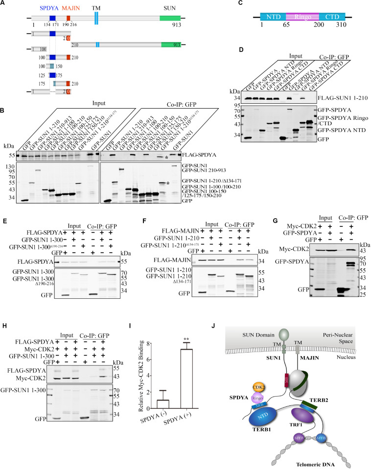
FIGURE 5.
SPDYA recruits CDK2 to SUN1. (A) Illustration of the SPDYA-binding site at SUN1. The SPDYA-binding site is shown in dark blue. TM: trans-membrane region. (B) The 134–171 Aa of SUN1 in the SPDYA binding site. *Indicating the band of GFP-SUN1 1–210 AaΔ 134– 171. (C) Illustration of the SPDYA structure. (D) The Ringo domain of SPDYA is the binding site of SUN1. (E) The interaction of SUN1 mutant with MAJIN-binding site deletion between SPDYA. (F) The interaction of SUN1 mutant with SPDYA-binding site deletion between MAJIN. (G,H) SPDYA interacts with CDK2 and recruits CDK2 to SUN1. (I) The histogram shows Myc-CDK2 binding in (H), data are presented as mean ± SEM with three repeats. **P < 0.01. (J) Illustration of the interaction of SUN1 with MAJIN and SPDYA-CDK2. The SPDYA Ringo domain binding site of SUN1 is shown in blue and name “SRB.”

