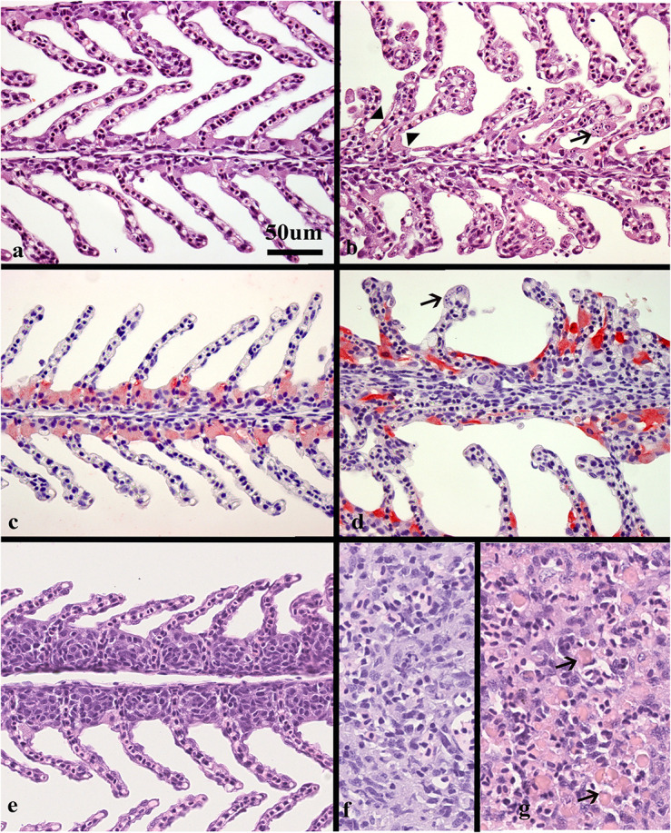
FIGURE 2.

Histological sections of gills and spleen from healthy and SGPV infected fish. (a) Sections of healthy gills with normal, thin lamellae stained with hematoxillin and eosin (HE), (b) gill from fish sampled from tank I at the start of the outbreak (M-I), gill epithelial apoptosis score of 2,5, SGPV Ct 18,36. Thickening of the lamellae due to apoptotic cells marked by arrow. Some of these cells appear to have the same staining properties as chloride cells. Note also some adhered lamellae (arrowheads). (c) Normal, thin lamellae, and chloride cells as detected by immunohistochemistry (light red) located in the filament epithelium. (d) gill from fish in tank II during the mortality peak (M-II group), gill epithelial apoptosis score of 1,5, SGPV Ct = 18,88. The chloride cells have a very intense staining and elongated shape compared to the healthy controls. The apoptotic cells (arrow) do not stain. (e) Gill epithelial hyperplasia (score 1), from fish in the L-I group with SGPV ct value of 27,9. (f) Normal spleen from the C-III group (g) spleen from the M-I group showing extensive hemophagocytosis (arrows).
