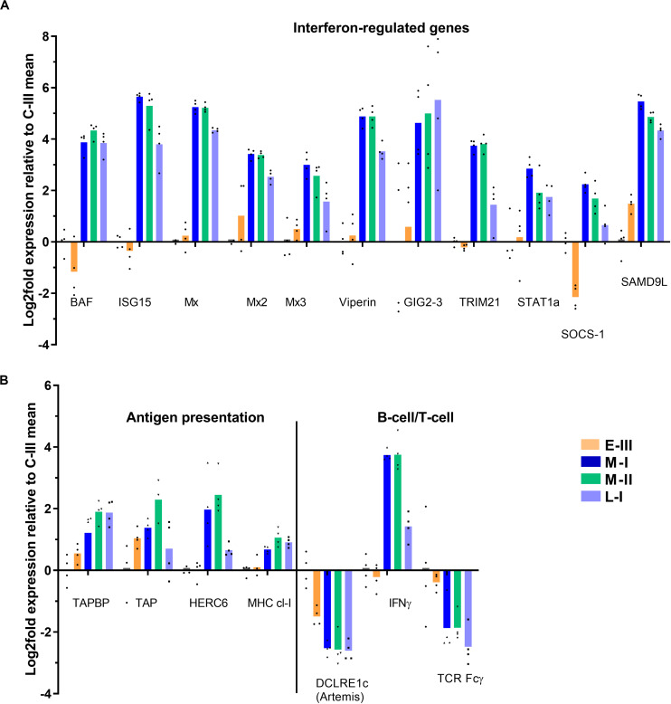
FIGURE 4.
Gene expression reflecting immune responses in gills from Atlantic salmon with SGPVD. Microarray data shown as log2 relative expression relative to control (C-III) mean expression. Mean values (bars) and individual values (dots) are shown. Bar colors: Orange (E-III, early infection), dark blue (M-I, peak infection/clinical disease), green (M-II, peak infection/clinical disease), and light blue (L-I, late infection/resolving disease). (A) Selected interferon-stimulated genes. BAF; Barrier of autointegration factor, ISG15; interferon stimulated gene 15, Mx; Myxovirus resistance genes, TRIM; Tripartite Motif protein, STAT; signal transducer and activator of transcription, SOCS; Suppressor of cytokine signaling, and SAMD9L; sterile alpha motif domain-containing 9-like protein. (B) Genes involved in antigen presentation and lymphocyte function. TAP; Transporter associated with Antigen Processing; TAPBP; TAP-Binding Protein, HERC; Homologous to E6-AP carboxyl-terminus (HECT)- and Regulator of Chromosome Condensation-1 protein (RCC)-domain containing protein, MHC; multihistocompatibility complex, DCLRE1c; DNA cross-link repair 1C/”Artemis”, IFN; Interferon, and TCR; T-cell receptor.

