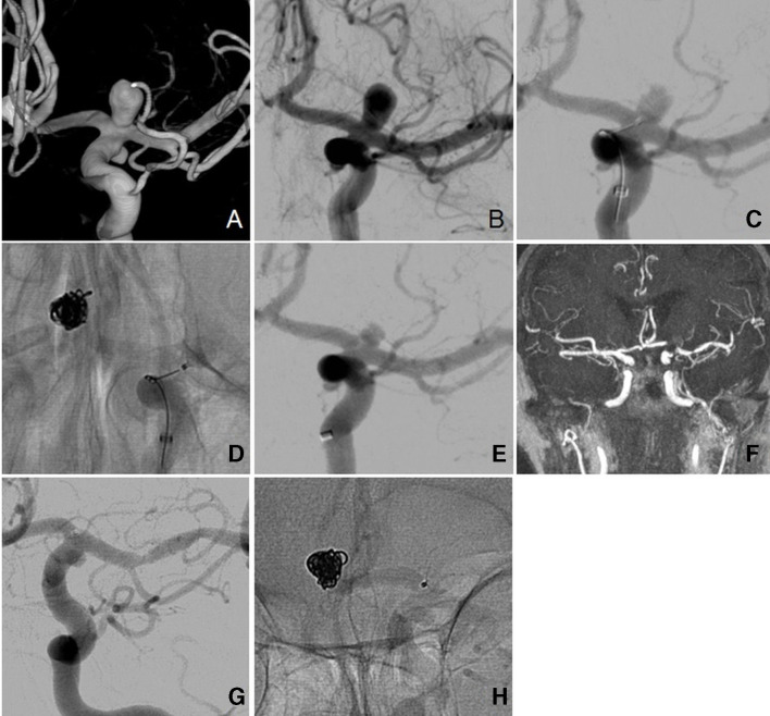
Figure 3.
Case 1. (A) Three-dimensional reconstruction of the internal carotid artery aneurysm. (B) Angiographic view of untreated aneurysm. (C) Microcatheter at aneurysm neck. (D) 7 mm Contour Neurovascular System deployed within aneurysm. (E) Early angiographic view showing signs of stasis. (F) Six-month follow-up MRI showing complete occlusion, although with artefact. (G) Six-month follow-up angiogram showing complete occlusion and flow diversion. (H) Angiographic view showing device remains in good position.

