Figure 2.

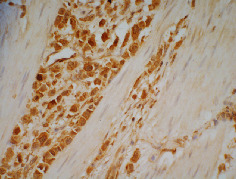
TIMP-1 positive staining. The membrane or cytoplasm of gastric cancer cells was stained brown. Original magnification, ×400.

TIMP-1 positive staining. The membrane or cytoplasm of gastric cancer cells was stained brown. Original magnification, ×400.