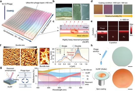
Figure 1.

a) Schematic illustration of colorimetric sensor with corresponding Ge thickness and relative humidity with M‐13 phage coating. b) SEM image showing phage‐coated porous Ge layer of cross‐sectional (left) and top view SEM image (right). Scale bar is 100 nm. c) Schematic showing highly lossy resonant promoter (HLRP) for resonance enhancement. d) Color images of colorimetric sensor corresponding to different humidity levels. e) Absorption intensity distributions of phage‐coated HLRP, Si, and Au, respectively. f) AFM images and thickness level profiling showing bundle size change by humidity conditions. Scale bar is 5 µm. g) Schematic illustration of resonant enhanced color reflection mechanism with phage layer swelling and reflectance spectra showing enhanced absorption and dip shift. h) Schematic of spin‐coating method and sample images onto 2‐inch wafer before (top) and after (bottom) phage spin‐coating. Scale bar is 1 cm.
