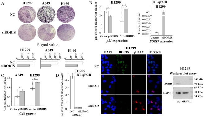
Figure 2.
BORIS promotes NSCLC cell proliferation. (A) BORIS knockdown suppressed colony formation of NSCLC cells. Crystal violet was used to stain the cells in upper panels. The bottom graphs present the signal difference in the upper panels. (B) Expression of p21 regulated by BORIS detected by RT-qPCR. The overexpression efficiency of pBORIS was assessed via RT-qPCR analysis (right panel). (C) BORIS overexpression promoted cell proliferation of A549 and H1299 cells measured by MTT assay. (D) Knockdown of BORIS by either siRNA-1 or siRNA-2 induced DNA damage efficiently measured by immunofluorescence stain of γH2AX (Red). BORIS proteins were detected by the primary BORIS antibody (FITC). The right and left panels presented transfection efficiencies of BORIS siRNA by western blot and RT-qPCR. *P<0.05; ***P<0.001. A549/DDP, DDP resistant A549 cell line; BORIS, Brother of Regulator of Imprinted Sites; NSCLC, non-small cell lung cancer; OD, optical density; RT-q, reverse transcription-quantitative; si, small interfering; nc, negative siRNA control.

