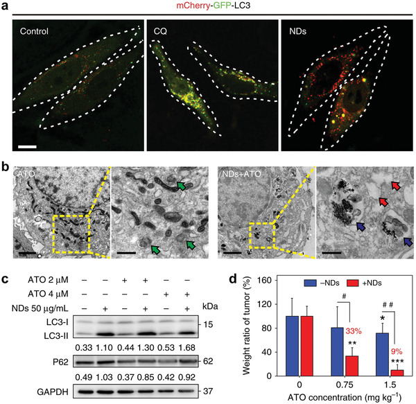
Figure 11.

Nanodiamond as an autophagy inhibitor for allosterically enhancing the chemotherapeutic effect of arsenic trioxide. a) Confocal fluorescence images of cancer cells transfected with mCherry‐GFP‐LC3 after incubation with chloroquine (CQ) or nanodiamonds (NDs) for 48 h (yellow, autophagosomes; red, autolysosomes). Scale bar, 10 µm. b) Bio‐TEM images of cancer cells treated with single arsenic trioxide (ATO) or the mixture of NDs and ATO. Scale bars, 1 µm for left low magnification images, and 500 nm for right high magnification images. Green arrows indicate structures of autophagosomes induced by ATO, while blue arrows indicate autolysosome containing NDs induced by NDs/ATO mixture. Additionally, red arrows point to vacuoles inside cells. c) Immunoblot analyses revealing the expressions of autophagy‐related proteins LC3 and p62 in cancer cells. Glyceraldehyde 3‐phosphate dehydrogenase (GAPDH) was used as a loading control. d) Weight ratio of tumors harvested from mice after different treatments. **P < 0.01, ***P < 0.001, significantly different from normal saline; #P < 0.05, ##P < 0.01, significantly different from ATO. Reproduced with permission.[ 42 ] Copyright 2018, Springer Nature.
