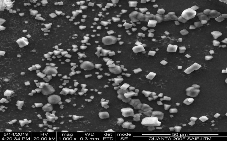
Fig. (3).
The scanning electron micrographs of sorafenib entrapped sodium selenite nanoparticles. The nanoparticles were formulated by using 0.5% w/v of tripolyphosphate through solvent evaporation method. The figure is self-exemplary of particles, which are discrete, crystal like structure with few are nano sized particles and most of the particles are micron sized particles.

