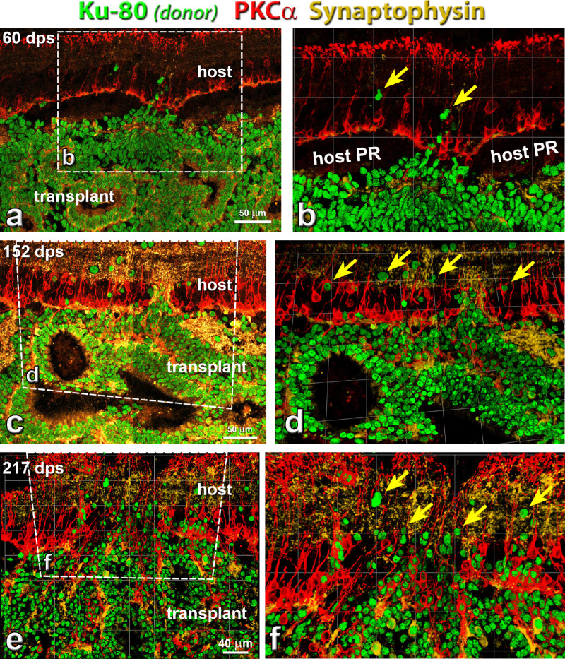
Figure 4.
Rod bipolar cells (PKCα) development in transplant, and connectivity between host and donor cells at different time points. Combination of label for PKCα (protein kinase C α; rod bipolar cells, red), Ku-80 (human nuclei, green) and synaptophysin (membrane protein of human synaptic vesicles, gold). (a, b) Around 60 days post surgery (dps), transplants started to form rosettes. A few donor cells migrated into host retina (yellow arrows). PKCα and Synaptophysin staining was found in the transplant but not very strong. (c, d) Around 152 dps, more donor cells migrated into host retina (yellow arrows). PKCα and Synaptophysin staining increased in transplant. (e, f) At 217 dps, more donor cells migrated into host retina than at 152 dps (yellow arrows) (transplant #8, see Table 3). Extensive synaptic connectivity (synaptophysin) was found between donor and host cells in the host IPL. Scale bars = 50 µm. (a, c); 40 µm (b, d, f) are enlargement of (a, c, and e). For abbreviations, see Table 5.

