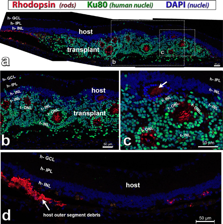
Figure 5.
Rhodopsin staining (red) in hESC-retina transplant (green nuclei) to nude RCS rat, 9.7 months after surgery (see also Supplementary Fig. S1). This rat (transplant #4, see Table 3) had good responses in the SC. Combination of rhodopsin (rods, red), Ku80 (human nuclei, green) and DAPI (nuclei, blue). (a) Overview. The transplant has developed photoreceptors in rosettes with outer segments (red). There seems to be some host rod photoreceptor rescue close to the transplant (left side in a). Donor nuclei are migrating into the host retina in some places. (b, c) enlargements of (a). Note that there is also a rosette of remaining host photoreceptors (only blue DAPI stain) in (c) (white arrow). (d) Host retina outside transplant, with outer segment debris on the left, and few scattered degenerated host rod cell bodies on the right. Scale bars = 50 µm. DAPI = 4’,6-Diamidino-2-phenylindole. See Table 5 for definitions of labels.

