Figure 6.
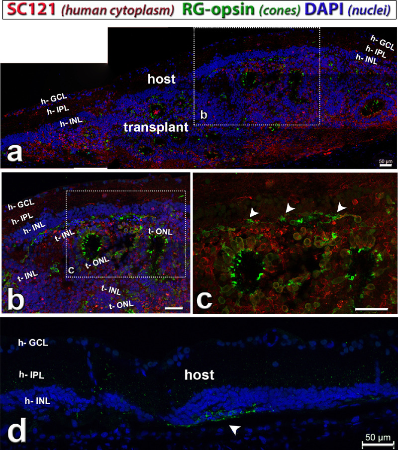
Cone photoreceptors (red–green [RG] opsin, green) in hESC-retina transplant (red cytoplasmic label) to nude RCS rat, 8.6 months after surgery (see also Supplementary Fig. S1). This rat (transplant #5) had good responses in the SC. Combination of SC121 (human cytoplasm, red), red–green opsin (cones, green) and DAPI (nuclei, blue). (a) Overview. The transplant developed photoreceptors in rosettes with outer segments (green). In the boxed area (enlarged in b, c) there were remaining host cone photoreceptors (arrow heads in c), but without outer segments. Donor processes in host IPL (h). (d) Host retina outside transplant depicting area with few degenerating host cones without outer segments (green, arrow head). Scale bars = 50 µm. DAPI = 4’,6-Diamidino-2-phenylindole. See Table 5 for definitions of labels.

