Figure 7.
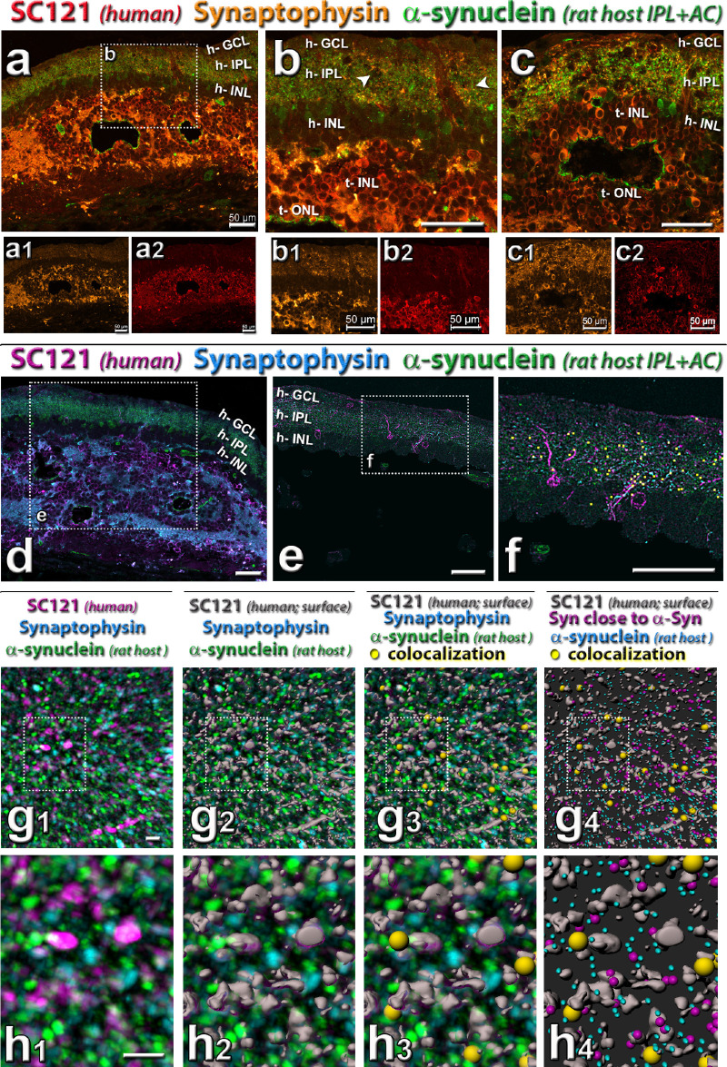
Triple staining for synaptophysin in combination with donor label SC121 and host cell marker (see also Supplementary Figs. S2 and S3). (a–b) Combination of SC121 (red), synaptophysin (gold), and rat-specific α-synuclein (green). Note the mixing of SC121, synaptophysin and α-synuclein in host IPL. (a, b, d–h) Same transplant as in Figure 5. Arrowheads in (b, e) point to areas of increased synaptophysin immunoreactivity and SC121 processes in the host IPL (h-IPL). (c) Same transplant as in Figure 6, showing transplant–host interface. (d) Combination of SC121 (magenta), synaptophysin (turquoise), and rat-specific α-synuclein (green): area adjacent to transplant shown in (a), with box showing area in (e). (e–h) Imaris software analysis of colocalization (closeness to) between host-specific α-synuclein, synaptophysin, and transplant label SC-121. (e) Mask applied to select mostly host retina. (f) Overlay of yellow squares shows colocalization of all three labels in the host IPL. (g, h) Enlarged three-dimensional rendering of detail in host IPL (all of the same area and magnification). The meaning of colors is indicated above the panels. (g1, h1) cleaned up label after thresholding. (g2, h2) SC121 staining rendered three-dimensional (grey) with Imaris surface function. (g3, h3) overlay of yellow dots showing triple colocalization. (g4, h4) Turquoise puncta indicate α-synuclein (host), purple puncta show synaptophysin close to α-synuclein, gray shows SC-121 surface; yellow dots show triple colocalization. Underlying image removed. Scale bars = 50 µm (a–f), 4 µm (g, h). See Table 5 for definitions of labels.

