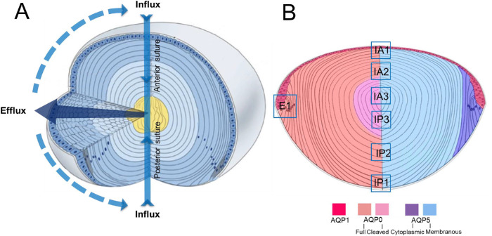
Figure 1.
Schematic diagrams illustrating lens structure and function and the distribution of AQPs in different regions of the lens. (A) Three-dimensional diagram of the lens showing ion and water fluxes coming into the lens core (yellow) via an extracellular route located at the anterior and posterior sutures (blue arrows). Ions and water cross fiber cell membranes before traveling via an intercellular pathway mediated by gap junction channels (dark blue arrow) to exit the lens at the equator. (B) Diagram of an axial section of the lens showing subcellular distributions of the lens water channels (AQP) in the different regions of the lens. AQP1 (red) is restricted to the membranes of the lens epithelium. AQP0 (left) is found in the membranes of lens fiber cells across all areas of the lens, but in the core of the lens, the C-terminal tail is cleaved. AQP5 (right) is also found throughout all regions of the lens, but in the epithelial (not shown) and peripheral differentiating fiber (purple) cells, it is associated with the cytoplasm. In deeper regions of the outer cortex, AQP5 becomes associated with the plasma membrane (blue), and this labeling extends into the lens core. In this study, we focused on the subcellular distributions of the three lens AQPs at the equator, which is associated with water efflux (E1), and the anterior (IA1, IA2, IA3) and posterior (IP1, IP2, IP3) poles, which mediate water influx. Adapted with permission from Shi Y, Barton K, De Maria A, Petrash JM, Shiels A, Bassnett S. The stratified syncytium of the vertebrate lens. J Cell Sci. 2009;122:1607–1615.

