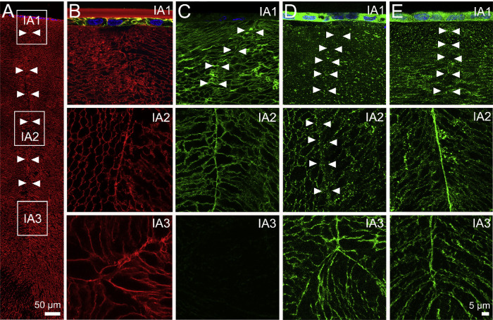
Figure 7.
Subcellular localization of lens AQPs in the influx zone of rat lenses—results from the anterior pole. (A) Image montage of the anterior water influx zone taken from an axial section of a rat lens that was labeled with the membrane marker WGA (red) to highlight suture line (arrowheads). Boxes indicate the areas (IA1, IA2, and IA3) from which the higher-resolution images shown in B–E were taken to investigate the subcellular distribution for each lens AQP (green). (B) In lenses with cut zonules, AQP1 labeling was present only in the epithelial cells of IA1 and was absent from IA2 and IA3. (C) In lenses with cut zonules, AQP0 labeling was membranous and strongly labeled the suture in regions IA1 and IA2, but no labeling was observed from region IA3 in the lens core where the C-terminus of AQP0 protein is cleaved. (D) In lenses with zonules cut, AQP5 labeling was missing from the suture in regions IA1 and IA2, but labeling was present in the deeper IA3 region. (E) In lenses that were fixed in situ with their zonules attached, AQP5 labeling was still absent from the suture in the peripheral region IA1 but was present in regions IA2 and IA3.

