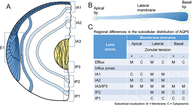
Figure 9.
Schematic representation of the subcellular localization of AQP5 in the efflux and influx zones of the rat lens in the presence and absence of zonular tension. (A) Epithelial cells (dark blue) that differentiate into fiber cells (blue) in the equatorial efflux zone (E1) are initially attached by their apical membrane domains to form the modiolus. As fiber cells detach from the modiolus, their apical and basal tips migrate along the epithelium and capsule, respectively, and their lateral membranes undergo massive elongation. This process of elongation continues until the apical and basal tips of fiber cells (light blue) from the opposing lens hemisphere meet to form the anterior and posterior sutures, respectively. As this process continues throughout life, newly differentiated secondary fiber cells internalize older mature fiber cells, which in turn internalize the primary fiber cells (yellow) laid down during embryonic development. Boxes represent the regions in the efflux and influx zones where the subcellular localization of AQP5 was measured. (B) Schematic representation of a fiber cell depicting its three specific membrane domains consisting of the apical tip, lateral membranes, and basal tip. (C) Table summarizing the regional differences of AQP5 subcellular localization observed in the efflux and influx zones in the presence and absence of zonular tension.

