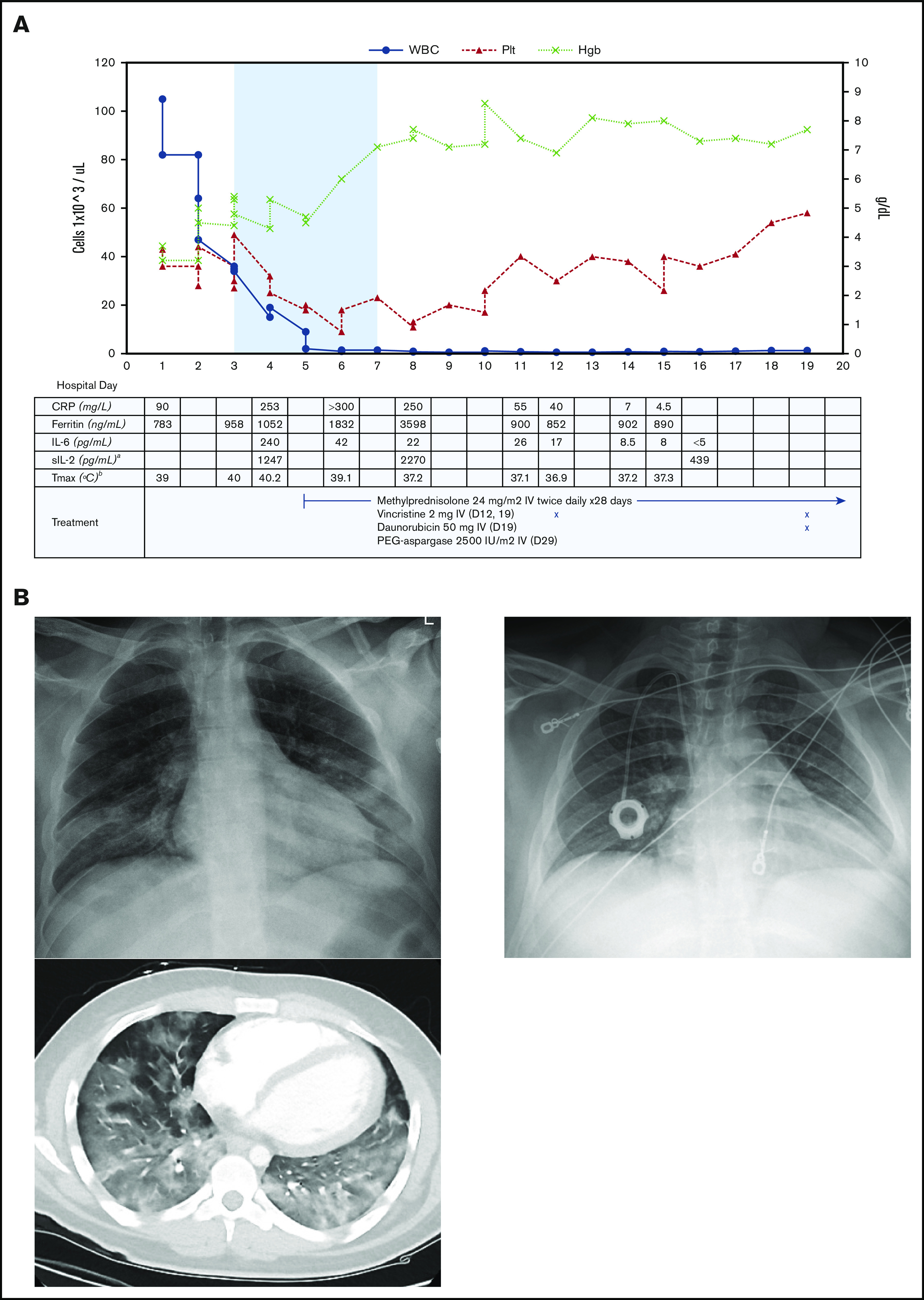
Figure 1.

Patient’s clinical and radiographic findings. (A) Cell counts and inflammatory markers throughout the patient’s hospital stay, from day of presentation to discharge. Top, The shaded area indicates time requiring mechanical ventilation. Left y-axis, WBC and platelet (Plt) counts in cells ×103 per microliter. Right y-axis, Hgb in grams per deciliter. Bottom, Patient’s antileukemic treatment included methylprednisolone, vincristine, and daunorubicin; timing staggered per clinical discretion. Systemic steroid therapy was started on HD 5, for a total course of 28 days. Vincristine was given on HD 12 and continued weekly for 4 doses. Daunorubicin was given on HD 19 and continued weekly for 4 doses. Treatment continued in the outpatient setting after hospital discharge, and additionally included 1 dose of polyethylene glycol (PEG)–aspargase. aIL-1β, IL-4, IL-5, IL-10, IL-12, IL-13, IL-17, interferon γ, and tumor necrosis factor α also tested without elevation in levels. bMaximum temperature (Tmax) recorded that calendar day. Normal reference values per reporting laboratory: C-reactive protein (CRP), 0-10 mg/L; ferritin, 30-400 ng/mL; IL-6, <5 pg/mL; sIL-2, <1033 pg/mL. (B) Chest radiograph and computed tomography scan at time of presentation (left), showing patchy consolidation and characteristic ground-glass pulmonary infiltrates throughout the lungs, predominant in the lower lobes. Chest radiograph 2 months later (right), showing resolution of consolidation.
