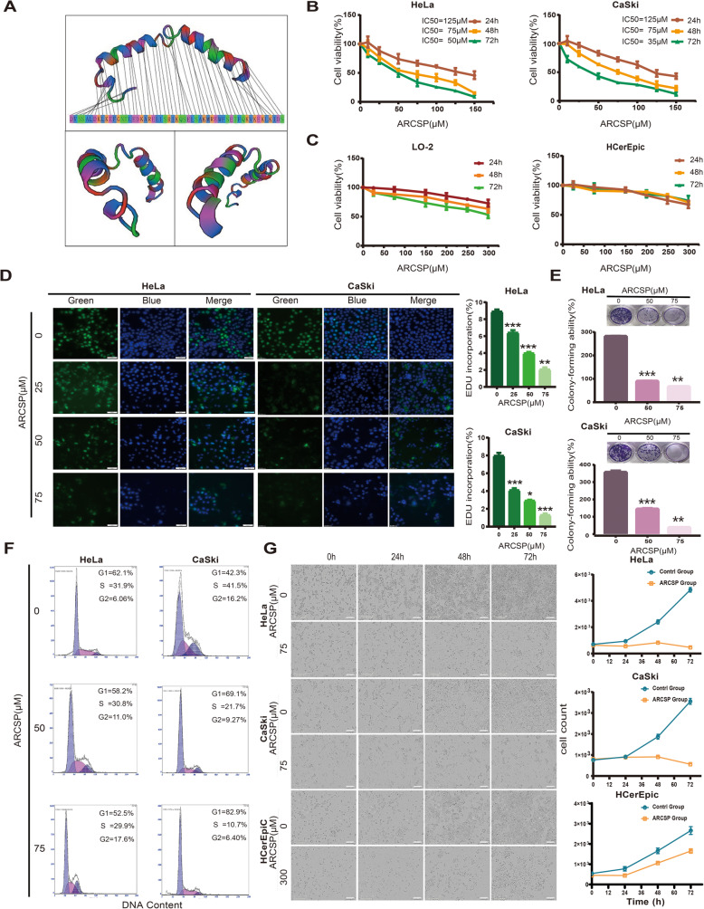
Fig. 1.
ARCSP inhibits the proliferation of cervical cancer cells and has low cytotoxicity in normal cells. a Model of the secondary structure of ARCSP. b Cervical cancer cell lines were treated with ARCSP (0–150 μM) for 24–72 h, and cell viability was measured by the CCK8 assay. c Normal cell lines (LO-2 and HCerEpic cells) were treated with ARCSP (0–300 μM) for 24–72 h, and cell viability was measured by the CCK8 assay. d Cells were treated with ARCSP (0–75 μM) for 48 h, and the proliferation of cells was measured by the EdU proliferation assay. Scale bar = 50 μm. The histograms show the quantified EdU incorporation data, which were calculated using ImageJ software. e Cells were treated with ARCSP (0–75 μM) for 14 days, and clone-forming ability was determined by the cell colony formation assay. The histograms show the quantified results of the colony formation assay, which were calculated using ImageJ software. f After 48 h of treatment with ARCSP (0–75 μM), cell cycle distribution was analyzed by FACS; the ratios of cells in G1, S, and G2/M phases are shown on the right. g After 72 h of continuous treatment with ARCSP (75 μM, 300 μM), the number of cells was significantly reduced. Scale bar = 300 μm. The data are expressed as the mean ± SD; *P < 0.05, **P < 0.01, ***P < 0.001. ns, not significant

