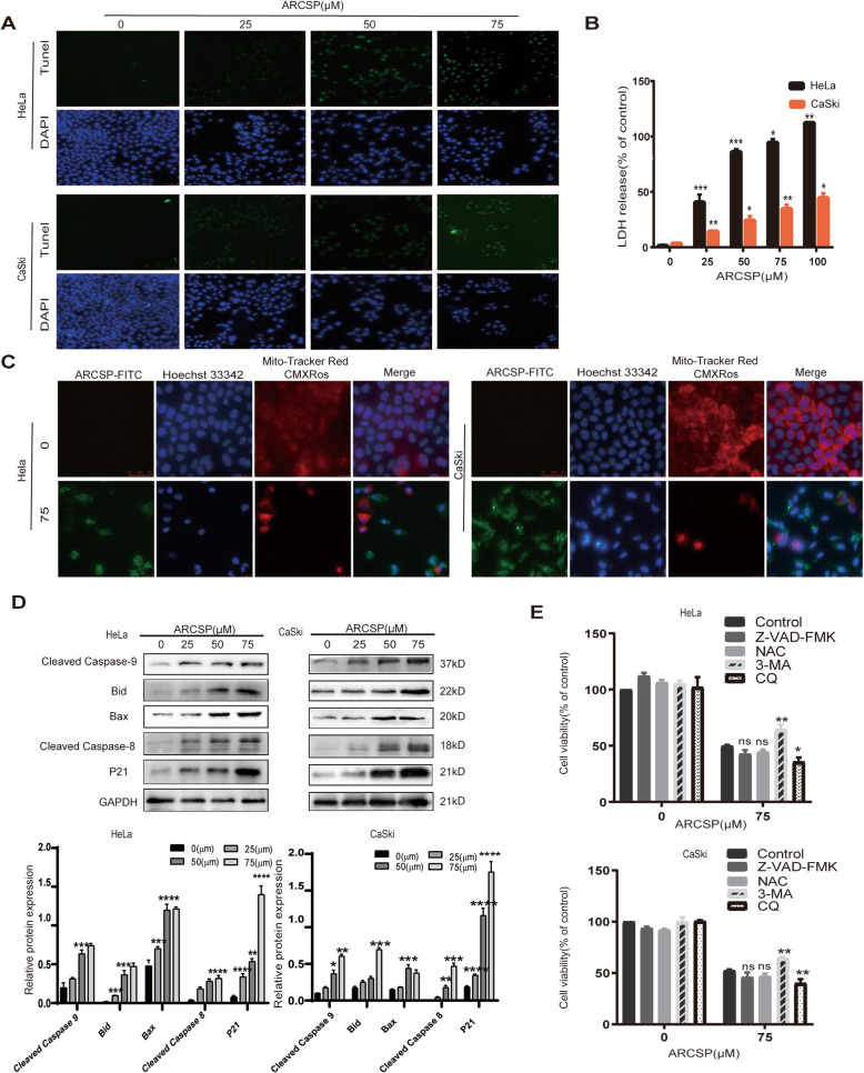
Fig. 2.
ARCSP induces apoptosis of cervical cancer cells. a Cells were treated with ARCSP (0–75 μM) for 48 h, and the apoptosis index was determined by the TUNEL assay. Scale bar = 50 μm. b Cells were treated with ARCSP (0–100 μM) for 48 h, and cell membrane damage was detected by the LDH release assay. c Cells were treated with ARCSP (75 μM) for 48 h, and the mitochondrial membrane potential was determined by MitoTracker Red staining, Hoechst 33342 (blue) was used to stain the nuclei. Scale bar = 25 μm. d Cells were treated with ARCSP (0–75 μM) for 48 h, and the expression levels of cleaved-caspase 9, Bid, Bax, cleaved-caspase 8, and P21 were detected. e Cells were cotreated with ARCSP and Z-VAD-FMK (5 μM), NAC (5 μM), 3-MA (20 μM) or CQ (20 μM) for 48 h, and cell viability was determined by the CCK8 assay. The data are expressed as the mean ± SD; *P < 0.05, **P < 0.01, ***P < 0.001. ns, not significant

