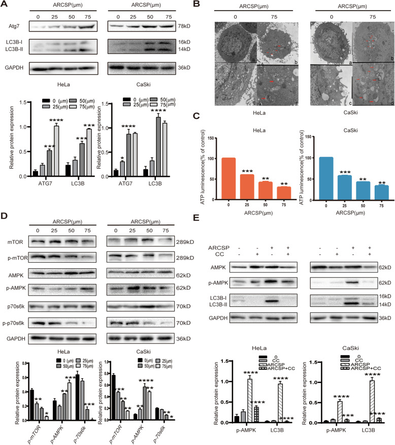
Fig. 3.
ARCSP induces autophagy, and the AMPK/mTOR signaling pathway may be involved in ARCSP-induced autophagy. a After HeLa and CaSki cells were treated with ARCSP (0–75 μM) for 48 h, we detected the expression of LC3B-I, LC3B-II, and Atg7 by Western blotting. b Cells were treated with or without ARCSP (75 μM) for 48 h. Autophagosomes were observed by transmission electron microscopy (a and b 5000×; c and d 15,000×). Scale bar = 0.1 μm. c Cells were treated with ARCSP (0–75 μM) for 48 h, and the ATP assay kit was used to detect intracellular ATP levels. d After cells were treated with ARCSP (0–75 μM) for 48 h, the levels of AMPK, p-AMPK, mTOR, p-mTOR, p70s6k, and p-p70s6k were detected by Western blotting. e The AMPK inhibitor CC (10 μM) was administered with ARCSP (75 μM) for 48 h, and the expression of LC3B-I, LC3B-II, AMPK, and p-AMPK was detected by Western blotting. The data are expressed as the mean ± SD; *P < 0.05, **P < 0.01, ***P < 0.001. ns, not significant

