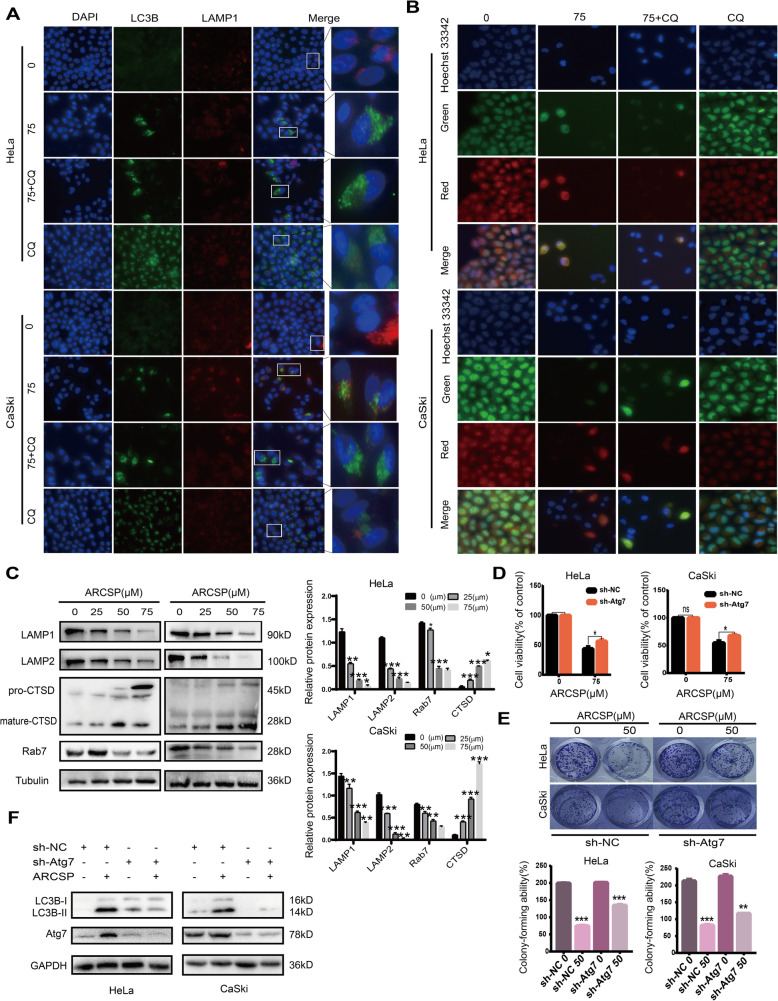
Fig. 6.
ARCSP may have a cytotoxic effect by affecting the function of lysosomes. a Cells were treated with ARCSP (75 μM) for 48 h, and the colocalization of LC3B (488 green) and LAMP1 (594 red) was assessed. DAPI (blue) was used to stain the nuclei, and the cells were photographed under a fluorescence microscope. Scale bar = 25 μm. b Cells were treated with ARCSP (75 μM) or CQ (20 μM) for 48 h, stained with AO for 10 min, Hoechst 33342 (blue) was used to stain the nuclei, and photographed under a fluorescence microscope. Scale bar = 25 μm. c Cells were treated with ARCSP (0–75 μM) for 48 h, and the levels of Rab7, LAMP1, LAMP2 and CTSD were detected by Western blotting. d Cells were transfected with sh-Atg7 and treated with ARCSP (75 μM) for 48 h, and cell viability was analyzed by the CCK8 assay. e Cells were transfected with sh-Atg7 and treated with ARCSP (50 μM) for 14 days, and the cell colony formation assay was used to evaluate cell proliferation. The histograms show the quantified results of the colony formation assay, which were calculated using ImageJ software. f Cells were treated with ARCSP (75 μM) for 48 h, and the expression of LC3B-I and LC3B-II was detected by Western blotting. The data are expressed as the mean ± SD; *P < 0.05, **P < 0.01, ***P < 0.001. ns, not significant

