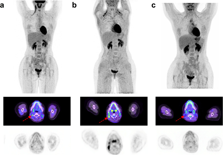
Fig. 2.
31-year-old female with Hodgkin Lymphoma. From top to bottom, Maximum intensity projection (MIP) PET, fused axial PET/CT and stand-alone PET images: a June 2018: Baseline PET showing stage 2 disease in the right neck. b January 2020: End-of-treatment PET considered non-diagnostic due to BAT activation in sites involved on baseline scan. c March 2020: follow-up PET examination. The patient received 40 mg of propranolol 1H prior to FDG injection. BAT activation is no longer visible and the patient is considered in complete metabolic response (Deauville score 2) with mediastinal uptake interpreted as thymic hyperplasia

