Fig. 2.
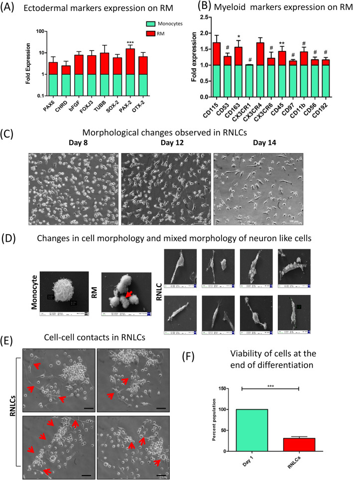
Characterization of lineage switch in reconditioned monocytes (RM) and their in vitro induction into retinal neuron-like cells (RNLCs). a Ectodermal markers were upregulated as compared to day 1 monocytes. b RM also contributed to express myeloid lineage markers (n = 4). c Morphology changes observed at × 20 upon induction with extrinsic retinal differentiation growth factors, where round and colony-forming RM formed long, elongated, round and axonal cells which we termed as retinal neuron-like cells (RNLCs) at day 14. d SEM images depicting a typical monocyte which further changes to dividing RM (indicated by red arrow) and eventually to RNLCs exhibiting mixed morphology of neuron-like cells. e The RNLCs also exhibited cell-cell contacts as indicated by red arrows. f Approximately 40% cells of the initial PBMCs were live and viable after induction at the culture endpoint as indicated by MTT assay

