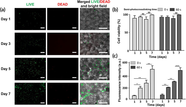
FIGURE 6.

In vitro biological activities of microporous hydrogels fabricated via annealing thermostable gelatin methacryloyl (GelMA) microgels. (a) Assessment of live (green) and dead (red) NIH/3T3 cells after 1, 3, 5, and 7 days of culture in three‐dimentional (3D) beaded GelMA scaffolds prepared from photo‐annealing semi‐photocrosslinked microgels (ultrabviolet [UV] exposure time = 60 s, intensity ~100 mW/cm2) via 120 s of UV light exposure at an intensity of ~10 mW/cm2. Brightfield images show the spreading of cells among GelMA microbeads. (b) Cell viability was measured based on the number of live cells divided by the total cell number in beaded GelMA scaffolds fabricated from semi‐photocrosslinked (UV exposure time = 60 s) or physically crosslinked (UV exposure time = 0 s) GelMA microgels photo‐annealed via 120 s of UV light exposure at an intensity of ~10 mW/cm2.(c) Metabolic activity of the cells embedded in the 3D beaded scaffolds as a function of incubation time, measured using the PrestoBlue® assay. Scale bars represent 200 μm
