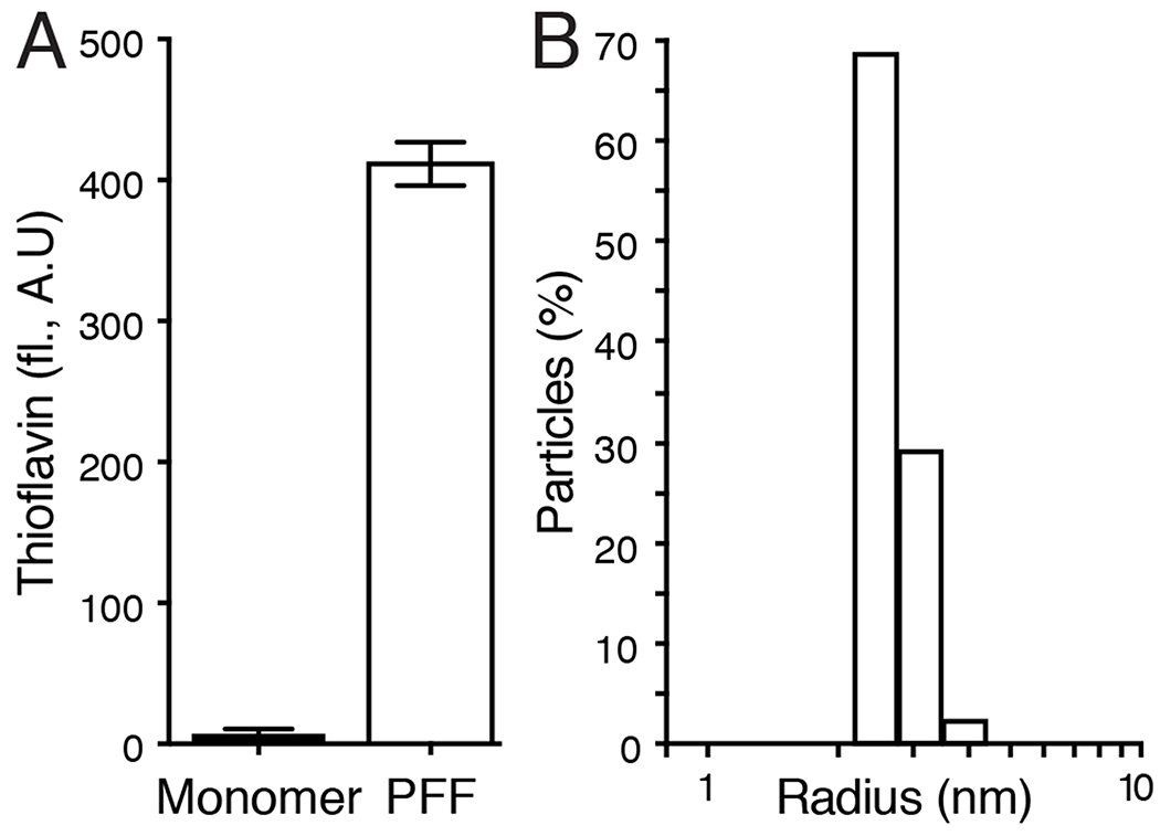
Figure 1. Biophysical characterization of α-Synuclein pre-formed fibrils.

(A) Thioflavin T fluorescence of monomeric α-synuclein and α-synuclein PFFs. Bars show mean +/− SEM fluorescence (arbitrary units) of 3 measurements. (B) Dynamic light scattering of α-synuclein PFFs. Bars show percent of particles at each hydrodynamic radius calculated by the time-dependent fluctuations in scattered light intensity using a Wyatt Technology DynaPro NanoStar instrument with DYNAMICS software.
