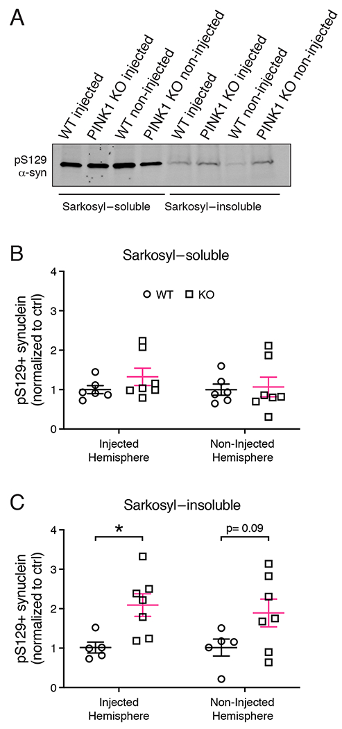
Figure 4. Levels of sarkosyl-soluble and insoluble α-synuclein in WT and PINK1 KO rat brains 4 weeks post PFF injection.

(A) pS129 α-synuclein western analysis of sarkosyl-soluble and insoluble fractions from whole brain lysates of PINK1 KO and WT rats 4 weeks post PFF injections. (B) Densitometric quantification of pS129 α-synuclein levels in the sarkosyl-soluble fraction. (C) Quantification of pS129 α-synuclein levels in the sarkosyl-insoluble fraction. Two-way ANOVA with Sidak’s multiple comparison showed a significant main effect of genotype F(1, 20)=11.21), *p = 0.0032. (n= 5 WT, 7 KO). Sidak’s multiple comparisons shows significantly increased pS129 α-synuclein in PINK1 KO rats in the injected hemisphere (*p = 0.0332), but not in the non-injected hemisphere (p = 0.09). Data were normal according to the Kolmogorov-Smirnov normality test. Error bars represent mean +/− SEM.
