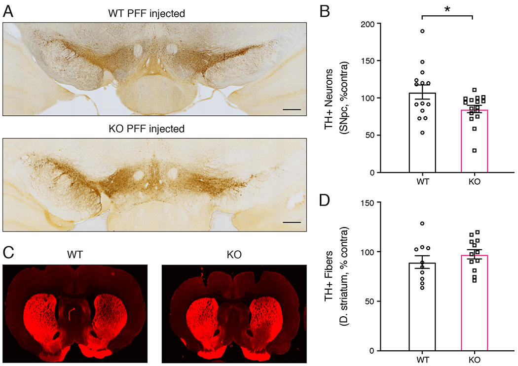
Figure 5. PFF induced neurodegeneration in PINK1 KO rats.

(A) Dopaminergic neurons analyzed by tyrosine hydroxylase (TH) immunohistochemistry of coronal brain sections WT and PINK1 KO rats 4 weeks post PFF injection. The region of the substantia nigra is shown. The injected hemisphere is on the right. Scale bars are 500 microns. (B) Quantification of TH+ neurons in the SNpc of WT and PINK1 KO rats by unbiased stereology. PFF injection caused a significant loss of dopaminergic neurons in PINK1 KO rats compared to WT rats (n= 14 WT, 16 KO) *p = 0.0464, unpaired t-test with Welch’s correction, t(19.71)= 1.125. (C) TH immunofluorescence of the dorsal striatum of WT and PINK1 KO rats 4 weeks post PFF injection. Data were normal according to the Kolmogorov-Smirnov normality test and the Shapiro Wilks test. (D) Quantification of TH immunofluorescence intensity in the dorsal striatum shows no differences between WT and PINK1 KO rats 4 weeks post PFF injection.
