Figure 1.

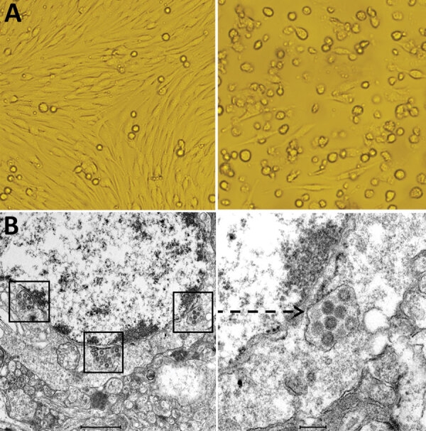
Cytopathogenic effect and electron microscopic morphology of baby hamster kidney 21 (BHK-21) cells infected with phlebovirus, China. A) Left panel shows morphology of BHK-21 cells before inoculation with strain SXWX1813-2; right panel shows morphology 3 days after inoculation (original magnification ×200). BHK-21 cells infected with SXWX1813-2 showed reduced adherence and a large number of rounded and exfoliated cells. B) Left panel shows the viral morphology of SXWX1813–2 on ultrathin slices (scale bar 1μm); right panel shows the enlarged viral particle (indicated by arrow) (scale bar 200nm).
