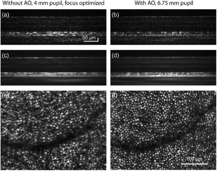
Fig. 6.
Cross-sectional and en face view of retina without and with adaptive optics. Single and averaged B-scans of retina at 7 deg. temporal to fovea are shown without (a,c) and with (b,d) adaptive optics. Corresponding en face images at COST layer are shown in (e) and (f). Separate scale bars are indicated for the B-scans and the en face images.

