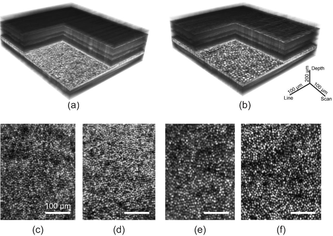
Fig. 7.
High-resolution imaging of retinal structure with AO line-scan spectral domain OCT and line-scan ophthalmoscope. Three dimensional rendering of AO-OCT volumes acquired at 2 and 4 deg temporal eccentricity are shown in (a) and (b). The corresponding en face images at the COST layer show cone photoreceptors in (d) and (f). LSO en face images are shown in (c) and (e). Scale bars for the volumes are indicated and are equal to 100 µm for the en face images.

