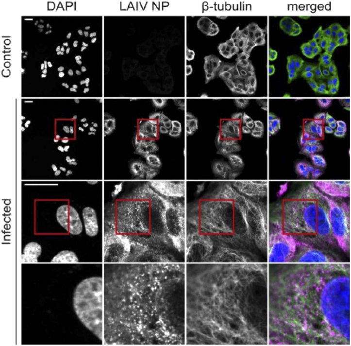
Fig. 1.
Non-expanded A549 cells infected with live attenuated influenza vaccine (LAIV). Cells were fixed 9 hours post infection and images recorded on a confocal microscope. Immunostaining was used to fluorescently label LAIV nucleoprotein (NP, magenta) and microtubules (green) whereas DAPI staining was used for labelling the nuclei (blue). Scalebar 25 µm. Size of images in bottom row 25 × 25 µm.

